
Cardiology Case #23
Primary Author: Dr Alastair Robertson; Co-Authors: Dr Hywel James and David Law
Background:
A male in his 40s presents to ED with several days of dizzy episodes with exertion, and associated palpitations. He has also had some intermittent chest tightness.
Physical examination is unremarkable and his observations are within normal limits.
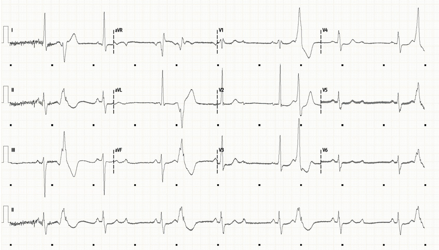
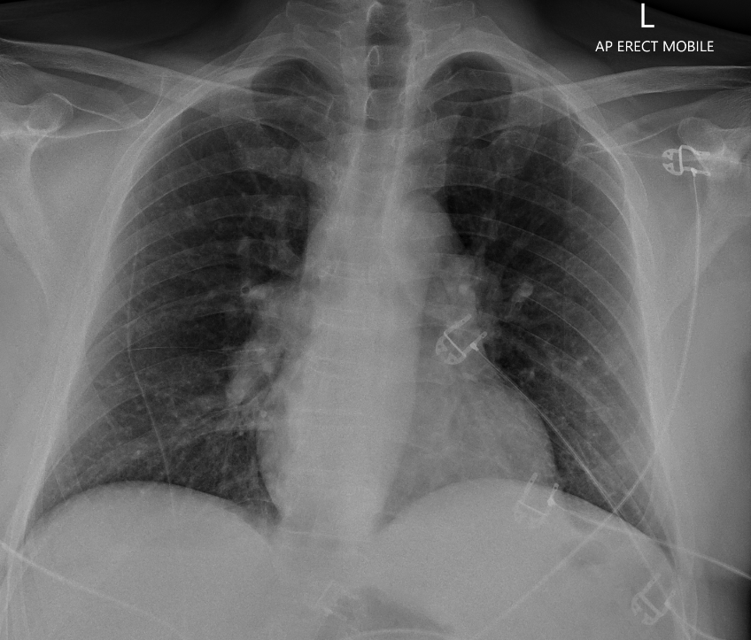
ECG and CXR are shown below
Initial Investigations:
This is an interesting ECG:
There is what looks like ventricular trigemini, with a PVC after every two normal sinus beats.
However, if you look closely (at the rhythm strip in particular) you will see that there is also a heart block. After each PVC there is a P-QRS-T, and then another P-wave which is not followed by a QRS.
This is a 2nd degree (Mobitz type 2) AV block.
Otherwise, there is some borderline left axis deviation with LAFB
No significant ischaemic changes noted
This ECG is concerning for high-grade AV block in the setting of pre-syncopal symptoms.
Chest XR shows clear lung fields, and a normal heart size. Interestingly the hilar regions appear bulky bilaterally however.
Clinical Synthesis:
Below is a parasternal long-axis view from this patient obtained in the emergency department. The echo features in combination with the information above gives a cardiology ‘spot diagnosis’ for a condition which is rare to pick up in ED.
What do you think of the echo, and do you know the diagnosis?
Basic POCUS: interpretation
The information from the PLAX clip is:
The initial impression is that LV function is impaired. There is moderate LV impairment, with an EF of around 40%, and the LV looks dilated.
More interestingly there is marked hypokinesis of both the proximal anterior septum (top), as well as the inferolateral wall (bottom - convention in echo nomenclature is to call this the inferolateral wall, but in ED it is still often called the posterior wall as it the one affected by a posterior MI). The apex is working more normally.
Thus there is segmental impairment, however this does not seem to follow any particular vascular territory (anterior septum is supplied by the LAD, but it would be very unusual to have proximal LAD or left main occlusion, with the distal septum looking normal. Additionally the inferolateral (posterior) wall is generally supplied by the RCA or the LCx.
There is also an unusual appearance of thinning of the proximal septum where it meets the anterior part of the aortic root.
In summary there appears to be a dilated cardiomyopathy with non-territorial segmental impairment, particular involving a thinned proximal anterior septum. This, combined with a diagnosis of conduction delay on ECG and bulky hilum on CXR is very suggestive of sarcoidosis.
Using our systemic review approach in Basic POCUS; there is no significant pericardial effusion, RV and aorta appear of normal size, whilst the left atrium appears dilated (which would be expected in the setting of dilated cardiomyopathy causing increased LA pressures).
Case Progress:
The patient was kept on telemetry to ensure he did not develop malignant arrhythmia. Troponins were normal. Further history revealed a previous diagnosis of likely sarcoid.
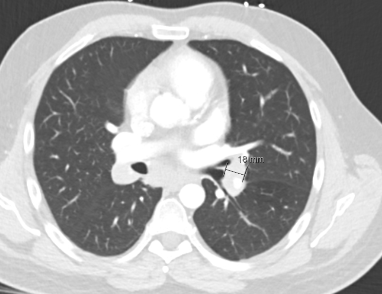
CT chest (slice shown below) revealed extensive bilateral pulmonary hilarity, subcarinal, and mediastinal lymphadenopathy as well as multifocal pulmonary nodules which would be consistent with pulmonary sarcoid.
FOCUS on Cardiac Sarcoid
Sarcoidosis, commonly shortened to just sarcoid, is a systemic, immune-mediated inflammatory disease characterized by the formation of non-caseating granulomas in affected organs. While it can involve virtually any organ system, it has a strong predilection for the thoracic cavity.
Is most commonly effects young-to-middle aged patients (typically 20-50yrs), and the most common presenting feature, particularly to ED is pulmonary disease.
General & Extracardiac Presentations
Pulmonary (>90% of cases): Progressive dyspnea, chronic non-productive cough, chest discomfort, and bilateral hilar lymphadenopathy on chest radiography.
Systemic / Constitutional: Low-grade fever, fatigue, weight loss, night sweats.
Cutaneous: Erythema nodosum (tender red nodules on shins), lupus pernio (indurated, violaceous plaques on the face/nose), or maculopapular lesions within old scars.
Ocular: Anterior/posterior uveitis (presenting as red eye, photophobia, or visual changes).
Renal/Metabolic: hypercalacemia or hypercalciuria.
Cardiac Sarcoid presentations:
Cardiac presentations can be very varied and mimic other pathologies, but are essentially an inflammatory process causing a myocarditis. Infiltrative granulomas in the ventricular walls classically presents with either conduction delays/heart block, or heart failure symptoms due to cardiomyopathy.
Conduction delays due to inflammation typically around the proximal septum involving the conduction system. Look for unexpected prolonged PR interval, bundle-branch block, or 2nd degree AV block in young patients.
LV impairment causing failure symptoms, fluid overload, exertional dyspnoea or decreased exercise tolerance. Can progress to severe cardiomyopathy with associated valvular disease.
Right heart failure due to either RV involvement, or due to increased pulmonary pressures from pulmonary sarcoid disease.
Malignant arrhythmia - cardiac sarcoid patients are at increased risk of sudden cardiac death
Definitive Diagnosis relies on either histological diagnosis from biopsy (of granuloma/thoracic lymphadenopathy, or cutaneous lesions), or if specific features are seen on cardiac MRI/PET. Supportive features can include XR/CT findings, echo findings, raised inflammatory markers, hypercalcaemia, and consistent cutaneous features.
Intermediate POCUS: Sarcoidosis on echo
Echo features of cardiac sarcoidosis to look for include:
Non-territorial regional wall motion abnormalities: hypokinesis which does not fit a particular vascular territory.
Dilated cardiomyopathy is often the end result of persistent cardiac inflammation.
Focal thinning of the basal interventricular septum - <7mm, or thinned compared to surrounding septum is common. This is the region through which the cardiac conduction system passes which explains why inflammation here can cause heart blocks. It is worth noting that sometimes acute myocardial inflammation can cause thickening of the myocardium which could be confused for hypertrophic cardiomyopathy (called paradoxical thickening, or ‘pseudotumour’.)
Review the above clip again. You can see the thinned basal septum and segmental LV impairment involving both anterior septum, and inferolateral (posterior) walls. If you look carefully you can see a slight region of lucency in the inferolateral (posterior) wall. This could be confused with a trace pericardial effusion, but is likely fibrosis/scarring in the myocardium secondary to sarcoidosis.
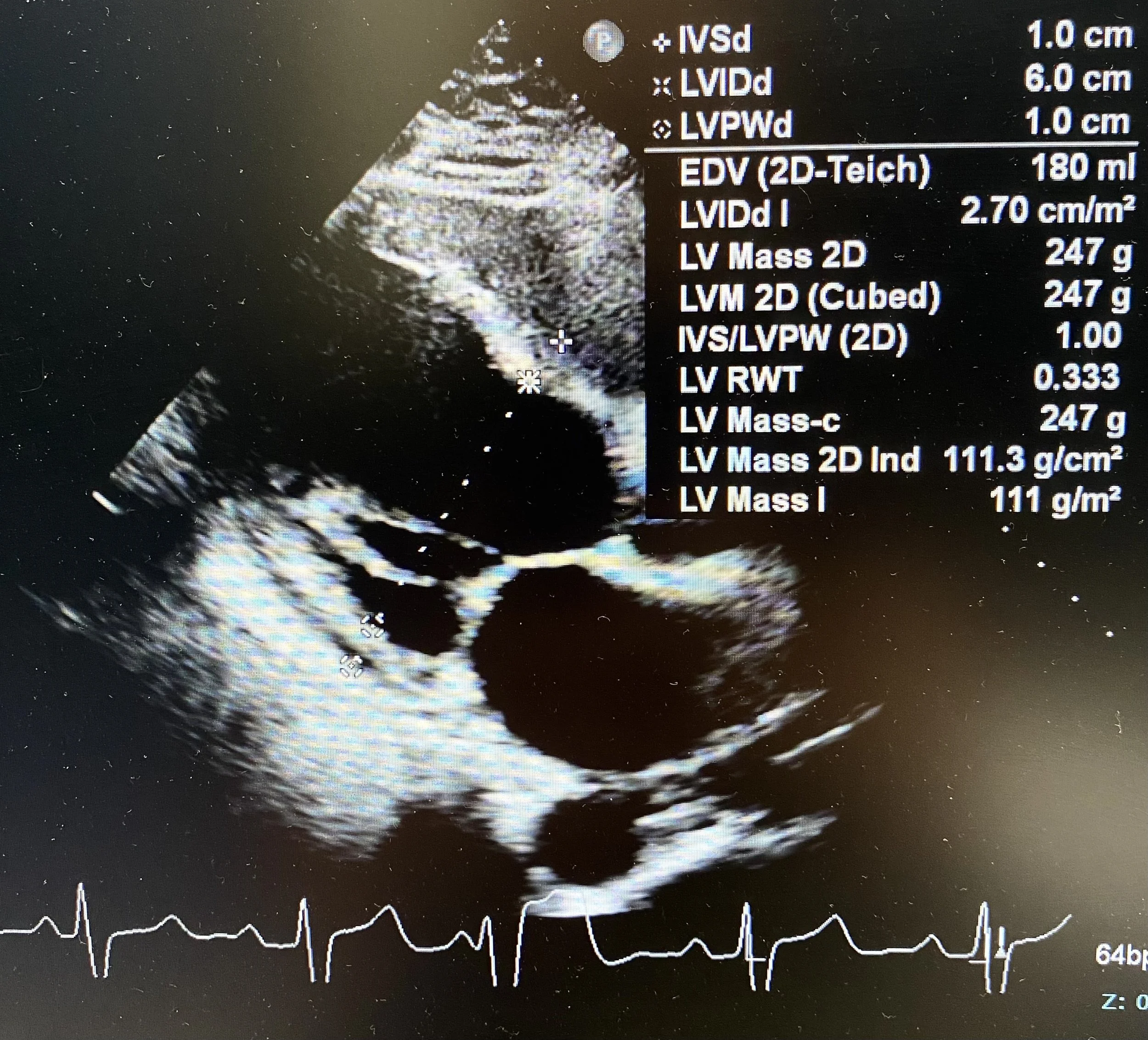
The LV is dilated (LVDd is 6.0cm), as confirmed by formal measurements below.
The parasternal SAX clip below also shows the non-territorial regional wall motion abnormalities.
Other echo features of cardiac sarcoid to look for include:
Impaired Global Longtitudinal Strain (GLS) - this is a more advanced echo technique to map out segmental impairment in LV function. GLS is often impaired before overall LV function so this can be a useful early marker of disease. Sarcoid typically shows a patchy, heterogenous loss of GLS. (This is different to cardiac amyloid which usually shows a symmetrical ‘bullseye’ or ‘apical sparing pattern’)
RV impairment - either due to inflammatory disease, or secondary to pulmonary sarcoidosis.
Ventricular Aneurysms - typically at the basal septum or posterior/lateral LV walls.
Pericardial disease- can present as pericarditis given this is an inflammatory condition, as well as pericardial effusion (tamponade has been reported but is rare).
Emergency Management of Cardiac Sarcoid:
Sarcoid is a relatively rare diagnosis and emergent management of the complications is generally by first principles.
AV Block:
Unstable bradycardia is managed along usual lines. Atropine is often ineffective if the block is infranodal so consider isoprenaline, adrenaline, or electrical pacing (transcutaneous or via pacing wire).
Malignant Arrhythmia:
Cardiac arrest due to malignant VT can occur. Management is via usual ALS but VT may be resistant to anti-arrythmics such as amiodacrone or lidocaine. Advanced strategies such as overdrive pacing or mechanical supports e.g. VA ECMO may be required.
LV failure:
As for other cardiomyopathy attempt to optimise fluid status, and consider adding inotropes if the patient is in frank cardiogenic shock.
Respiratory complications:
Consider the following as causes of respiratory distress in the setting of sarcoidosis.
Pulmonary HTN due to pulmonary disease causing RV failure, or acute PE given that sarcoid is a hyper-coagulable inflammatory state.
LV impairment causing functional mitral regurgitation can cause pulmonary oedema.
Secondary infection - often opportunistic such as PJP, TB, or fungal infection. Aspergillomas can cause pulmonary haemorrhage and severe haemoptysis.
Pulmonary disease progression due to severe alveolar granulomatous inflammation can cause respiratory failure.
Pneumothorax - in late stage disease pleural granulomas or associated bullae can break down to cause pneumothorax.
Upper airway obstruction due to laryngeal or supraglottic disease. The stridulous patient may require you to secure the airway emergently. Expect a difficult airway.
Hypercalcaemia
Is common in sarcoidosis due to macrophages in granulomas releasing enzymes which drive GI absorption of calcium. Features of hypercalcaemia are abdominal pain, constipation, bone pain, confusion/psychosis. ECG may show a short QT interval with osborne/J waves and a risk of ventricular dysrhythmia/VF.
Management is with IV fluids as long as the patients cardiac function will tolerate them. Unlike most other causes of hypercalcaemia bisphosphonates and calcitonin have reduced utility and are usually avoided. The first line treatment for hypercalcaemia in sarcoid is systemic corticosteroids.
Immunosuppression
High dose corticosteroids such as IV methylprednisolone may be required in the ED for acute fulminant presentations of sarcoidosis in conjunction with specialty advice. Otherwise oral prednisolone (e.g. 1mg/kg) is often the initial management.
Long-term management:
Many patients with isolated pulmonary sarcoid will go into spontaneous remission, but immunosuppression is generally required in cases of progressive pulmonary disease or multi-system disease. Agents may include corticosteroids, methotrexate, azathioprine, or biologics (such as Infliximab or Adalimumab).
Heart failure is treated along usual lines with fluid optimisation and a combination of ACE-inhibitors, beta-blockers, and diuretics.
ICD insertion is recommended for higher-risk patients with cardiac sarcoid (in particular those with documented malignant arrhythmia, LV EF <35% or high-grade AV block which requires PPM insertion anyway)
Ultimately cardiac transplantation may be required for end-stage refractory cardiac sarcoidosis.
