
Cardiology Case #24
Primary Author: Dr Alastair Robertson; Co-Authors: Dr Hywel James and David Law
Background:
A 78 year old male is brought into ED by ambulance from his home with a chief complaint of worsening bilateral lower limb swelling and erythema. He has been treated in the community with two courses of oral antibiotics for lower limb cellulitis with no improvement.
Notable background includes COPD (still smokes), chronic lower limb swelling, and recurrent DVT/PE including a breakthrough PE on warfarin. For this he is on long-term therapeutic dose enoxaparin.
On examination he had bibasal crackles on chest auscultation, a mildly distended abdomen, and bilateral erythema and swelling to both lower legs.
RR 24, Sats 94% on ambient air, HR 97/min, BP 88/66, temp 36.5
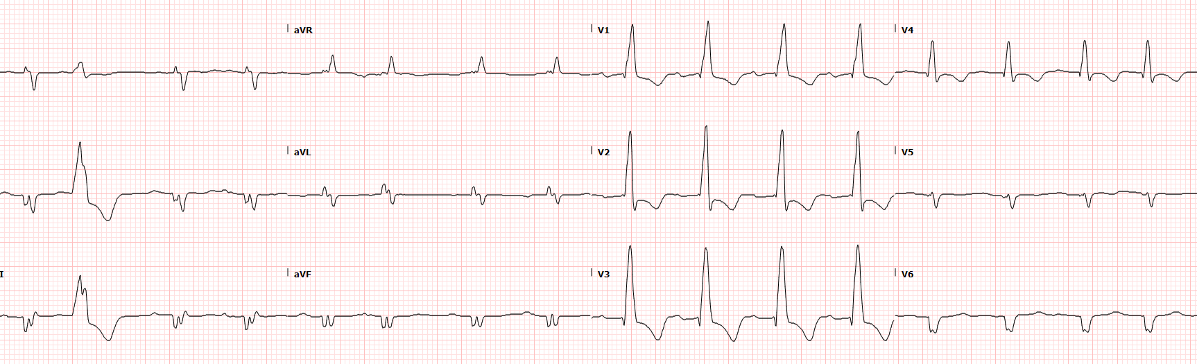
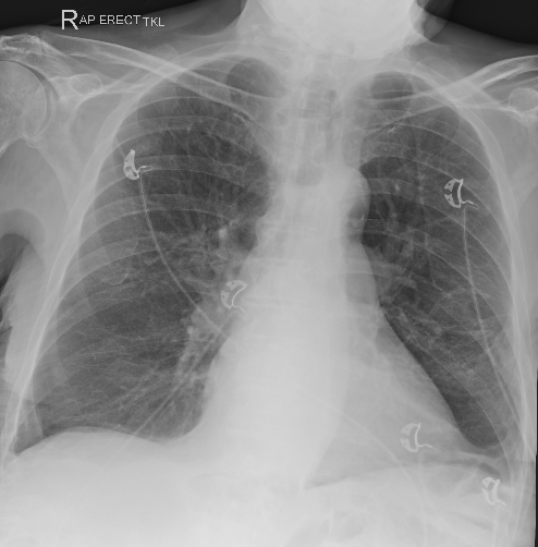
Above is his ECG and Chest XR, what is your interpretation, and how could POCUS be useful in this patient?
Initial Investigations:
This is an important ECG to recognise.
The rate is largely regular and there appear to be p-waves but this may be atrial fibrillation with a rate of around 90/min. The PR is borderline prolonged at just over 200ms.
The axis is unusual. The QRS is negative in leads I, II, and aVL. This is an extreme axis, often called a “Northwest Axis”. Differentials to consider when you see extreme axis deviation are:
Limb lead misplacement (most commonly RA and LL leads swapped)
Ventricular arrythmia - in particular VT
Severe right ventricular hypertrophy or strain
Hyperkalaemia which can cause multiple bizarre ECG changes
Dextrocardia or Situs Inversus
The QRS is broad at about 130ms meeting criteria for bundle branch block. This pattern is a RIGHT Bundle Branch Block.
There are marked T-wave inversions with ST depression, particularly in the precordial leads V1-4. Whilst ischaemia is a possibility, this pattern is characteristic of right ventricular strain.
This ECG suggests right heart pathology, when you see this pattern consider RV strain secondary to acute PE.
Chest XR shows some probable increased pulmonary vascular markings as well as blunting of the costophrenic angles suggesting small pleural effusions. Heart size appears enlarged although this is an AP film. There is no pneumothorax or focal consolidation.
Clinical Synthesis:
So although this patient is reasonably stable, they are hypoxic and hypotensive. Bilateral lower limb cellulitis is rarely a correct diagnosis. Venous stasis dermatitis from fluid overload (e.g. due to heart failure) is more likely. This patient has clear features of RV strain on his ECG and is very high risk for both DVT and PE despite his long-term anti-coagulation. PE needs to be excluded, but can POCUS help us differentiate between acute and chronic right heart issues?
Below are the Parastenal Long Axis (PLAX), and an Parasternal Short Axis (PSAX)
What do you think is happening?
Can you identify left and right ventricles?
Basic POCUS: global assessment
This is quite a spectacular echo and may be hard to orientate yourself. The top clip (PLAX) has the LV at the bottom of the screen, with a grossly dilated right ventricle at the top of the clip. You can see the LVOT and aortic valve leaving the left ventricle. The thick band running across the right ventricle is the moderator band. Note how the RV forms the apex of the heart rather than the LV.
The short axis clip (bottom) has the LV located at the bottom of the screen, with the severely dilated right ventricle above. If you focus on the interventricular septum you can see the flattened “D-shaped” septum during systole. Looking more closely you can see that although the septum becomes more rounded during diastole, it it still flattened, so we have both systolic AND diastolic septal flattening here.
The last clip here is a subcostal 4-chamber view. The RV is at the top of the screen and you can see that it is nearly twice the size of the LV. The tricuspid valve is clearly seen, with its associated papillary muscles in the RV. The small cavity LV is at the bottom of the screen and this clip nicely shows the abnormal movement of the interventricular septum, bowing into the LV rather than towards the RV as you would expect. This suggests that RV pressures are higher than LV pressures throughout the cardiac cycle.
Case Progress:
Our initial views above have shown a spectacularly dilated right ventricle, and a patient who is on the precipice of catastrophic RV failure. Given she sheer size and dysfunction of this RV it was concluded that most of his problems were chronic, as it is not possible to generate such a severely dilated RV with an acute insult.
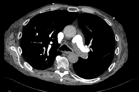
CTPA was performed to exclude new clot burden, and it showed a large PE in the left pulmonary artery, but this was unchanged from his last CT a few months prior (see slice below).
FOCUS on: Right Heart Failure
The right ventricle is easily overlooked yet it is crucial to have a functioning RV to maintain cardiac output. Remember that as a normal circulation is a closed system, the RV stroke volume must equal that of the LV, so RV impairment can rapidly impact your systemic perfusion.
The RV can be tricky to visualise well on echo despite being the most anterior structure. However it is important to consider RV failure in any unwell patient, and echo assessment of RV function (or RV to pulmonary arterial ‘coupling’) forms a key part of assessing the shocked patient (particularly using the 4-interface shock model - more of this in future posts).
Echo in Right Heart Failure
Assessment of the right heart can fall into several categories.
Here is what to look for on echo:
Chamber Dimensions & Geometry
RV: LV basal diameter ratio - (measured from an RV-focussed apical 4-chamber) is important. A ratio of >0.6 shows RV dilation, whilst a ratio of >1.0 marks severe RV dilation, and the RV usually takes over the apex.
RV basal diameter - again measured in an RV focussed A4C at end diastole - a diameter of >4.1cm is abnormal. A diameter >4.5cm is moderately dilated, and a diameter >5cm is severely dilated.
An RV controlling the apex of the heart is abnormal and suggests significant RV overload, usually representing chronic RV strain.
Interventricular septal flattening suggests RV impairment and likely raised pulmonary pressures. It is best assessed using a PSAX view at the mid-papillary level (LV mid-cavity). Flattening of the septum:
In systole only - suggests RV pressure overload
In diastole only - suggests RV volume overload
In both systole & diastole - suggests both pressure AND volume overload
A further measurement that can be done is the LV Eccentricity Index (EI) which is obtained by measuring both the shortest and the longest LV diameter in short axis (PSAX) at end systole. A normal ratio is 1 (a circular LV), however if the septum is flattened the LV will be elliptoid, and a ratio of >1.2 indicates septal flattening and elevated RV pressures.
Systolic Function Metrics
Measurement of TAPSE (Tricuspid Annular Plane Systolic Excursion) using M-mode in the A4C essentially gives a quantitative measure of RV shortening along its long plane. This is a reasonable gross surrogate for RV function because a healthy RV contracts primarily in this longitudinal plane with less radial contraction as compared to the LV. A normal TAPSE is considered over 18mm, however there are caveats with measuring TAPSE and it should form part of the overall assessment. It is often unreliable in the setting of previous cardiac surgery, RV infarction, and in severe TR.
TAPSE:PAP ratio can be another useful measurement to gauge if the RV is coping with its afterload by dividing the TAPSE (in mm), by the pulmonary artery pressure (in mmHg). A normal value will be >0.5. A value of less than 0.3 suggests uncoupling (or dysfunction) of the RV to PA interface, indicating RV failure.
There are many more advanced ways to assess RV function, which may be incorporated into a formal echo, but are less commonly utilised in the POCUS setting. These may include:
RV outflow doppler - PW doppler placed in the RVOT (from a PSAX window) will classically show a shortened acceleration time (time to peak), as well as notching of the downslope if the pulmonary pressures are elevated. Notching is specific but not sensitive, so should raise concern for PHN if it is seen.
RV Strain Patterns - similar to LV strain, this measures contractility by tracking ultrasound points in the RV wall. An RV strain below 20% signifies RV impairment.
Fractional Area Change - which is performed by mapping the RV areas during the cardiac cycle (similar to EF). An area change of <35% signifies global RV dysfunction.
RV Tissue Doppler - similar to LV TDIs, a lateral TV annulus tissue doppler S’ below 9.5cm/s suggests impaired RV function.
Haemodynamics & Congestion
Apart from assessing RV size, this is one of the most important assessments to make.
Tricuspid Regurgitation Vmax
The velocity of the TR flow allows assessment of the pressure difference across the tricuspid valve. If you add the right atrial pressure to this, you can estimate the systolic pulmonary artery pressures (PASP).
Use CW doppler through the TR jet to estimated the pressures. RAP is typically between 5-12mmHg, but may be up to 15mm with a dilated and plethoric IVC.
Normal PASP is below 40mmHg, and may reach as high as 80-90mmHg in severe pulmonary hypertension. Remember that this value will fall as the right ventricle fails, so a normal or mildly elevated value is not necessarily reassuring.
IVC Assessment & Venous congestion
IVC diameter >2.1cm, or a plethoric IVC suggest venous congestion which is an outcome of right heart failure. Note that IVC assessment can be unreliable if the patient is on positive pressure ventilation, or has abdominal compartment syndrome. If the IVC is not dilated but venous congestion is suspected then further assessment using the VexUS protocol assessing hepatic or portal vein flow may be useful.
Intermediate POCUS
Below is an apical 4-chamber view showing the severely dilated right ventricle which control the cardiac apex, note the abnormal septal motion and poor RV function. Note the right atrium is also dilated - whilst it does not look large in comparison to the RV, relative to the LV size it is moderately to severely dilated.
See the TR colour jet on the subcostal 4-chamber view. The photo below shows the CW doppler signal through the TR jet showing a maximum velocity of 340cm/s which corresponds to a pressure gradient of 51mmHg (taken from an apical window).
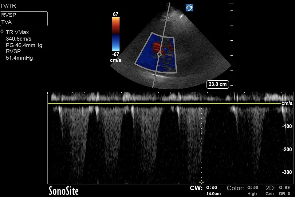
This patient’s IVC was dilated with minimal respiratory variation so RA pressure was estimated at 12-15mmHg making the PASP around 65mmHg. This is severely elevated.
Tips for assessing pulmonary pressures:
Try to find a window with alignment of the CW doppler with the TR jet. In ideal circumstances this is often from an apical 4-chamber, or an RV focussed view. Often a better trace can be obtained from sliding the probe medially towards a low parasternal position. This view will foreshorten the RV thus distorting its appearance but may yield a better TR trace.
If the apical windows are poor try to get alignment of the TR jet from a parasternal long window (tilt the probe inferiorly to bring in the tricuspid valve), or from a subcostal window (this may work in vertically orientated hearts such as in COPD).
The TR jet should be parabolic in shape, it is possible to confuse it with the aortic outflow jet which runs just next to it from the A4C window.
Increasing the CW doppler gains may help identify the parabolic shape.
If there is severe TR on colour doppler then the TR velocity will underestimate the actual pressure gradient.
After measuring the TR jet, add the estimated right atrial pressure (usually 5-12mmHg, based off the IVC size).
FOCUS on: Acute vs Chronic Right Heart Strain
Differentiating acute from chronic RV failure can be difficult. Not least because patients with chronic RV failure can (and do) develop acute issues such as PE. One key point to remember is that a previously normal (or ‘unconditioned’ RV) will struggle to generate pressures greater than about 60mmHg, so if the pulmonary pressures are severely elevated (>70-80mmHg) then there is an element of chronic pulmonary hypertension.
The following characteristics may help differentiate acute from chronic RV strain:
RV free wall thickness: in acute RV failure the wall thickness is usually <5mm. If it is greater than this then this may represent chronic hypertrophy. Thickened and prominent trabeculations and/or moderator band are an other indicator of chronic strain. Note how hypertorphied the RV walls and moderator band are in the clips above.
Degree of dilation: severe dilation (where the RV:LV ratio is above 1) or massive dilation (where the RV size dwarfs the LV, such as in this case) are characteristic of chronic RV stress. In acute PE the RV:LV ratio is unlikely to be greater than 1.0.
TR jet velocity: as mentioned above a velocity of >3.7-4.0m/s (corresponding to a pressure of >60-80) usually implies chronic remodelling. In acute PE the TR jet is usually <3.5m/s (or <55-60mmHg).
Regional wall motion: in acute PE you may see McConnell’s sign, which is RV lateral free wall hypokinesis with preserved or hyperdynamic contraction at the apex. The RV base may still be contracting in acute PE as well, leading to a preserved/normal TAPSE. In chronic RV failure you will tend to see global hypokinesis of a dilated RV with reduced TAPSE.
RVOT Doppler: as mentioned above a shortened acceleration time and RVOT notching are both seen in PHN. In acute PE the acceleration time is often <60ms with early systolic notching. In more chronic causes of PHN the notching is often later in the trace (so called ‘mid-systolic notching’).
Right Atrium: in acute causes the RA does not have time to dilate, so will be normal size, or mildly dilated. In chronic RV strain the RA dilates to decompress the RV, and may be severely dilated.
Case Progress:
This patient had chronic progressive RV failure, without evidence of new acute insult such as worsening PE. Evidence that this was chronic included:
Massive dilation of the RV with marked hypertrophy/thickening of the RV walls and a very prominent hypertrophied moderator band (as visible in the clips). The RV base measured 7cm!
Global hypokinesis of the right ventricle, lacking a McConnell’s sign, and the RV dominating the apex.
A severely dilated right atrium
He was admitted to hospital for management of his worsening RV failure which progressed to biventricular failure. After a complicated hospital stay he continued to deteriorate and was ultimately palliated.
FOCUS on: RV Failure & PHN Management
The causes of RV failure typically fall into three categories: increased afterload, impairment of contractility, or increased preload. When managing an unstable patient, it is important to differentiate between an acute pathology causing RV failure, and a patient with chronic pulmonary hypertension who is decompensating because of a different pathology such as sepsis.
Increased RV Afterload
Acute causes of increased RV aferload include:
Acute PE (massive/submassive) - PE will often cause haemodynamic collapse before it causes profound hypoxia.
Acute respiratory failure - such as ARDS, APO, or severe pneumonia causing hypoxic vasoconstriction.
Tension pneumothorax
Chronic causes are generally covered within the spectrum of Pulmonary Hypertension which are classified by the WSPH (World Society of Pulmonary Hypertension) into the following groups:
Group I - pulmonary arterial hypertension which can idiopathic/inheritied, caused by drugs (sympathomimetics), or secondary to neonatal PPHN.
Group II - left heart disease which can be caused by valvular issues (mitral/aortic), cardiomyopathy, or congenital causes such as shunts.
Group III - lung disease such as COPD, OSA, interstitial lung disease, or chronic high altitude.
Group IV - venous thromboembolism (called CTEPH) from chronic PEs with persisting clot burden. Other causes can include cancers, or arteritis.
Group V - multi-factorial or other causes e.g. haemolytic anaemia or metabolic/glycogen storage disorders.
Primary Myocardial Failure
Acute causes include RV infarction (from RCA occlusion), myocarditis (e.g. viral, immunotherapy), septic cardiomyopathy or biventricular Takotsubo, or RV stunning post cardiac surgery or cardiac arrest.
Chronic causes include chronic biventricular cardiomyopathy (which could be ischaemic, alcoholic, chronic Chagas disease etc.), infiltrative disease (amyloid, sarcoidosis, haemochromatosis), or ARVC (arrhythmogenic right ventricular cardiomyopathy).
Increased RV preload
Acute causes of this include:
Acute severe TR - which can be caused by endocarditis, or traumatic leaflet rupture (this can be caused by pacemaker wire removal).
Aggressive fluid resuscitation in a patient with pre-existing RV impairment.
Chronic causes include:
Tricuspid Regurgitation - functional from RV dilation, or organic.
Pulmonary Regurgitation - rarely seen as an acute issue but not uncommon after repair of congenital Tetralogy of Fallot.
Shunts - left-to-right shunts such as ASD, VSD, anomalous pulmonary venous return
Restrictive Causes
The final thing to be aware of is constrictive causes such as acute pericardial tamponade (usually acute), or a constrictive pericarditis (more chronic) as these will make the RV fail before the LV due to the lower pressures.
Management of RV failure in the ED
Acute RV failure is one of the most difficult things to manage acutely in ED.
Key management points are:
Treat the precipitating cause - If the patient has an acute PE, then prioritise thrombolysis or thrombectomy, for example. If the patient is septic, then prioritise antibiotics and source control. If they are in hypoxic respiratory failure then optimise their oxygenation and ventilation.
Following on from this, good resuscitation requires focussed supportive care:
1) Support RV perfusion pressure - the RV is perfused throughout the cardiac cycle so a target a higher blood pressure target to ensure this. A target MAP of >65-75, or a SBP equal to the RVSP may be required to avoid worseing RV ischaemia.
Noradrenaline is a reasonable first-line vasopressor to increase MAP due to relatively little effect of pulmonary vasculature. Vasopressin is also a reasonable agent for this.
Avoid pure vasoconstrictors such as phenylephrine as they may increase pulmonary vascular resistance (PVR) and RV afterload.
2) Optimise RV Preload - the RV is preload dependent when it is failing, but ED patients are more likely to be hypervolaemic than hypovolaemic. Excess venous and RV congestion elevates RV wall stress, decreases contractility, causes worsening TR from annular stretch, and increases adverse ventricular interdependence. Increased central venous pressure also increases capillary afterload decreasing end-organ perfusion.
in certain cases such as acute PE or pericardial tamponade small fluid boluses may improve RV filling and function but exercise extreme caution with IV fluids, and limit boluses to small volume (250ml) with close monitoring using serial echo.
As a general rule, cease any routine or maintenance fluids, they are far more likely to harm rather than benefit the patient.
Consider diuresis if there is an adequate blood pressure to support it, and evidence of venous congestion, with loop diuretics such as furosemide.
3) Support RV contractility - if RV contractility is poor then adding inotropy will help forward pulmonary flow but at the risk of increasing pulmonary vascular resistance. Reasonable agents may include:
Adrenaline - at low doses good inotropic effect which is relatively sparing of pulmonary vasculature, and it will not cause peripheral vasodilation.
Dobutamine is an effective inotrope for RV failure, but will cause peripheral vasodilation which may need to be countered with a peripheral vasopressor such as noradrenaline.
Milrinone is an excellent agent which causes pulmonary vasodilation coupled with increased inotropy. It’s profound vasodilatory effects and prolonged half-life can be problematic and it is usually reserved for more stable patients in the ICU setting.
4) Minimise RV afterload - this is achieved by lowering the pulmonary vascular resistance (PVR) as much as possible, thus reducing stress on the RV. Some of the agents listed above will aid with pulmonary vasodilation but some more specific strategies include:
Avoid hypoxia - aim for sats strictly >92-94% to avoid hypoxic pulmonary vasoconstriction elevating PVR.
Avoid acidosis or hypercarbia where possible as this will also increase PVR.
Use inhaled pulmonary vasodilators to reduce PVR. Common agents include inhaled nitric oxide (iNO) or inhaled prostacyclin. They can be delivered via high-flow nasal cannula, NIV, or via a ventilator circuit by various proprietary devices and have the benefit of pulmonary vasodilation without systemic vasodilation. Each has its own merits but choice of agent will likely be dictated by the availability in your centre.
5) Ventilation strategies - are complex in these patients. There is a high risk of haemodynamic collapse with induction for intubation so the risks and benefits need to be carefully considered. In general:
Resuscitate before any intubation attempt, and select agents/doses with the goal of minimising hypotension or apnoeic time during induction.
Avoid excessive PEEP which will reduce venous return where possible.
In general, use low tidal volumes (around 6ml/kg), and avoid high plateau pressures where possible to keep pulmonary capillary resistance low (with the caveat that resistance will also increase if driving pressures are too low).
Avoid hypercarbia, as mentioned above.
6) Serial Echo - to ensure that your interventions are working consider tracking:
RV to LV ratio, and interventricular septal movement - unlikely to change drastically but try to ensure things are not getting worse.
IVC size and collapsibility is easy to measure in serial exams and will give an insight into RV preload.
TR jet velocity (RVSP), or perhaps a TAPSE:PASP ratio to track improvement or deterioration of RV to PA uncoupling
LVOT VTI is a useful measure of systemic forward stroke volume (and thus RV stroke volume). In the setting of acute PE for example, a low LVOT VTI is a marker of obstructive shock.
7) Mechanical supports - for refractory cardiogenic shock rescue therapies could include dedicated RV supports (such as percutaneous RVADs) or VA ECMO which simultaneously offloads RV preload whist providing systemic organ perfusion.
