Cardiology Case #25
Primary Author: Dr Alastair Robertson; Co-Authors: Dr Hywel James and David Law
Background:
A man in his 70s is brought to ED by ambulance after a collapse whilst gardening with brief loss of consciousness. He is diaphoretic and nauseated but denies active chest pain.
He has a background of T2DM, hypertension and high cholesterol.
His observations are: RR 29, Sats 95% on air, HR 110, BP 106/66, temp 37.9.
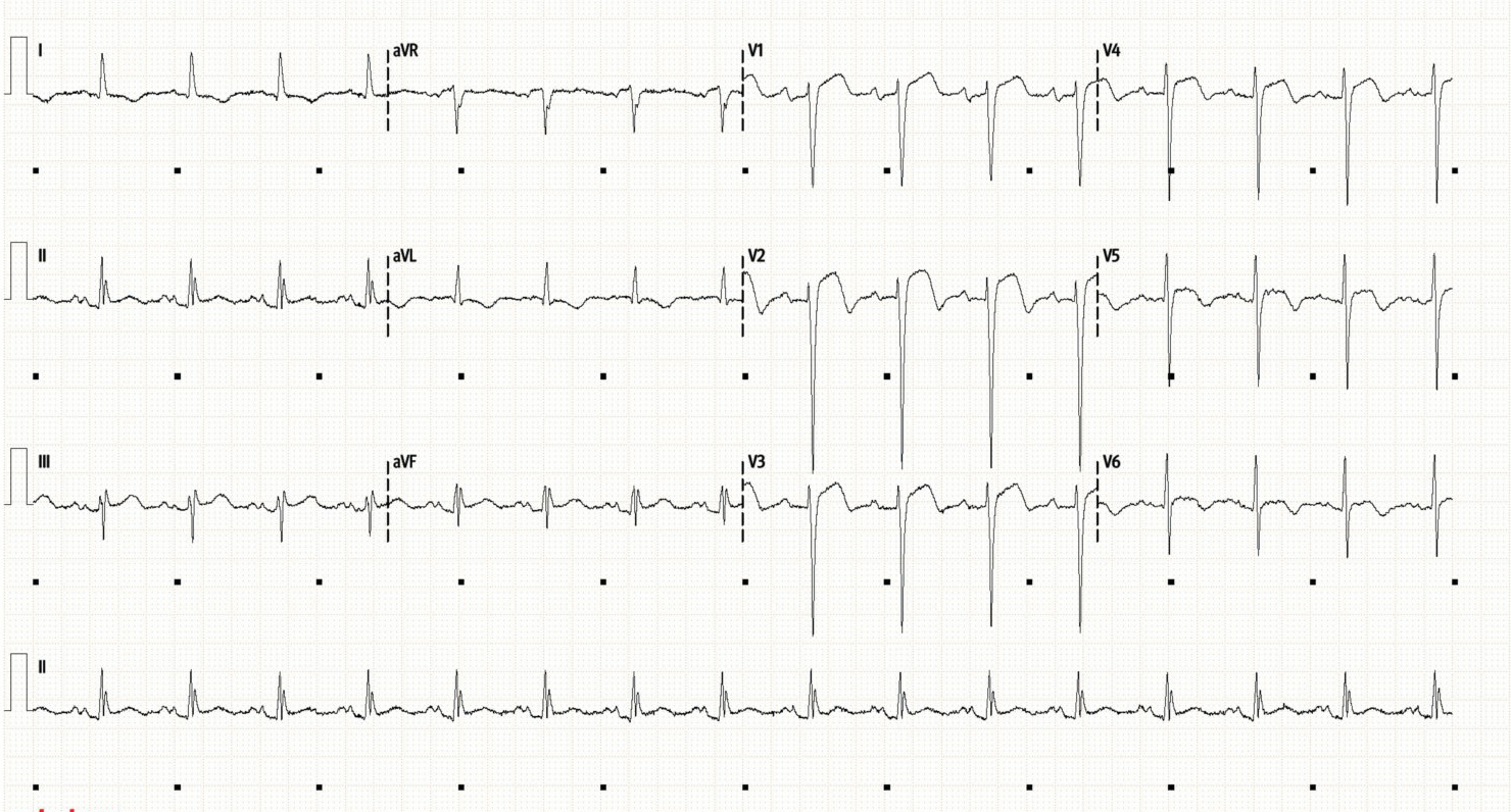
His initial ECG is below:
Initial Assessment:
This ECG was interpreted as an anterior STEMI
Sinus rhythm with a rate of around 100/min.
Anterior ST elevation, meeting STEMI criteria are seen in V2, V3, V4, and V5.
There is delayed R-wave progression, and reciprocal ST depression in the high lateral leads (I, aVL).
Cath lab was available when this patient presented so was activated immediately.
Basic POCUS
Initial echo in Resus while awaiting cath lab activation is shown below and beautifully illustrates how echo can correlate with ECG changes to confirm the diagnosis of STEMI with LAD occlusion.
PLAX (top), and PSAX (bottom)
Basic POCUS: interpretation
The top clip (PLAX) shows good function of the inferolateral (posterior) wall at the bottom of the screen which is usually supplied by the RCA. Anteriorly, the proximal septum is working (usually supplied by the left main or very proximal LAD), but beyond that you can see marked hypokinesis of the anterior septum extending into the apex.
The bottom clip (PSAX) shows the LV close to the apex. There is contraction inferiorly/posteriorly, but otherwise there is marked hypokinesis. Given most of the apex is supplied by the LAD this is strongly suggestive of an LAD occlusion.
LV function is likely moderately impaired given the apical involvement with a compensatory hyperdynamic LV base.
One can see how this combination of apical hypokinesis with compensatory hyperdynamic basal segments such as seen here could be confused with Takotsubo cardiomyopathy. This is why it is crucial to exclude LAD occlusion before making a diagnosis of Takotsubo. Stress cardiomyopathy must involve different vascular territories (which is NOT seen here) and is far less likely than MI in a male patient with cardiac risk factors.
Case Progress:
The patient proceeded to PCI where he was found to have severe LAD disease with long-segment plaque and two focal severe lesions which were successfully stented.
He was stable post-procedure and admitted to CCU.
Intermediate POCUS
Further assessment by echo was required when the patient subsequently became persistently hypotensive on CCU.
What differentials would you be considering?
Below is an apical 4-chamber view.
Intermediate POCUS - post-MI complications
In a patient in the acute MI period the differentials for haemodynamic deterioration are broad.
Important causes to consider are:
Ischaemic Complications:
LV free wall rupture, or septal rupture (VSD)
Valvular - papillary muscle rupture causing severe mitral regurgitation
Myocardial ischaemia causing LV impairment and cardiogenic shock
Mechanical obstruction such as dynamic LVOTO
RV failure (RV infarct, fluid overload, hypoxia from pulmonary oedema)
Electrical Complications:
AV block causing bradycardia/complete heart block
Tachyarrhythmias - VT, atrial fibrillation/flutter, SVT
Procedural Complications:
Stent occlusion or thrombosis, stent dissection, or microvascular obstruction
Pericardial tamponade/haemopericardium from coronary guidewire injury - this can cause a slow bleed which only become apparent hours after PCI.
Iatrogenic aortic dissection from guide wire injury
Occult Bleeding
Consider bleeding from arterial access sites, particularly if femoral as this can cause retroperitoneal haematoama.
GI bleeding from stress ulcer
Pharmacological
Anaphylaxis from iodinated contrast
Hypovolaemia from hyper-osmolar contrast agents or diuresis
Secondary to sedatives/analgesia used in cath lab, or newly started beta-blockers/diuretics.
Intermediate POCUS - interpretation
The A4C clip above shows that the LV function is reasonable (moderately impaired) with no large pericardial effusion or obvious free wall or septal rupture. You can see that the lateral wall, and the inferior (proximal) septum are contracting, whilst there is persisting apical hypokinesis in the LAD territory. RV size/function appears normal.
Of note this patient has a prominent/thickened proximal septum and you can also see a large mobile anterior mitral valve leaflet (left) flicking towards the mid-cavity. These features, combined with the apical impairment causing a hyperdynamic base, raised concern for dynamic left ventricular outflow tract obstruction (LVOTO).
So, how do we identify this on echo?
Intermediate POCUS: Dynamic LVOTO
We have talked about dynamic LVOTO previously. Look for a combination of predisposing factors and an acute precipitant.
Predisposing factors:
Small cavity LV
Hypertrophic proximal septum hypertrophy (HCM, hypertensive heart disease, ‘sigmoid’ septum in the elderly)
Alterations to or abnormal LV morphology (previous mitral/aortic valve replacement, ‘floppy’ mitral valve leaflets, post aortic stenosis repair)
Precipitating Events:
Hypovolaemia, Tachycardia, and/or peripheral vasodilation (e.g. septic, distributive, or hypovolaemic shock, anaesthetic drugs)
Hyperdynamic LV base (usually due to either LAD ischaemia or Takotsubo cardiomyopathy)
Excessive inotropy (iatrogenic e.g. adrenaline/dobutamine for shock, or sympathomimetic toxidromes)
Dynamic LVOTO on Echo:
The key to identifying LVOTO is noticing predisposing factors in the LV as well as a conducive haemodynamic state to create a high index of suspicion for it.
A hypotensive patient who is getting worse despite increasing inotropic support should always be a point to step back and consider dynamic LVOTO.
The key features for the diagnosis are:
1) Systolic Anterior Motion (SAM) of the Anterior Mitral Valve Leaflet in 2D
2) Aliasing (high velocity) flow through the LVOT
3) Mitral Regurgitation
4) Elevated LVOT Gradient
Dynamic LVOTO - SAM and 2D features
In most cases of dynamic LVOTO the LV (either the whole LV, or the LV base) can be seen to be hyperdynamic. This combined with features such as a small LV cavity and prominent or hypertrophied proximal septum should raise concern for the is phenomena. An LVEF >65%, complete obliteration of the LV cavity during systole, LVH (>15mm), or sigmoid septum are all risk factors.
A pathognomic feature of dynamic LVOTO is SAM, or systolic anterior movement of the anterior mitral valve leaflet. This is caused by high-velocity systolic blood flow through the LVOT pulling the closed mitral valve leaflet anteriorly due to the Venturi effect. It is therefore seen in mid-to-late systole. This has the dual effect of worsening the LVOT obstruction, as well as causing mitral regurigitation as this creates a defect in the mitral valve.
SAM can be hard to pick up in real time whilst scanning so is best seen by zooming on the mitral valve and then reviewing the clip one frame at a time, or viewing at slower speeds. It can be detected from both PLAX as well as A5C views. If using a PLAX view then M-mode can be used to track the mitral valve leaflets so see if the anterior leaflet moves anteriorly during systole (3-lead ECG tracing is useful to assess this).
Below is the zoomed clip of this patients mitral valve from an A5C view. See how the anterior (left hand) leaflet is pulled over into the septum during mid-to-late systole creating a dynamic LVOTO. You can also see the thickened proximal septum and hyperdynamic LV base which is contributing to this phenomenon.
If SAM with septal contact occurs and is present for >30% of systole this strongly correlates with haemodynamically significant LVOT gradients (>30-50mmHg).
Dynamic LVOTO - Aliasing Flow
Using colour doppler place the colour box over the LVOT and aortic valve. This can be done best in either the PLAX or from an Apical 5-chamber view. Set the colour scale reasonably high (to around 70 cm/s) and look for high-velocity flow through the region which will appear as either light yellow or light blue/white. Aliasing flow occurring in the LVOT rather than around the aortic valve leaflet tips suggests a high gradient through the LVOT rather than aortic stenosis. Sometimes mid-cavity outflow obstruction will also be seen with colour doppler.
The A5C view below shows aliasing flow through the LVOT.
Dynamic LVOTO - Mitral Regurgitation
Dynamic LVOTO with SAM creates a coaptation defect in the mitral valve, and thus the combination of blood not being able to leave the ventricle via the outflow tract - with an incompetent mitral valve - causes significant mitral regurgitation.
Due to the mechanics this creates a characteristic eccentric posteriorly- and laterally-directed MR jet, which occurs in late systole and is usually severe. Significant dynamic LVOTO is unlikely if there is no MR.
As will be discussed in the next section, the MR jet is often detected when putting CW doppler through the LVOT to measure the gradient. Learn to differentiate the parabolic, and high velocity MR signal, from the dynamic LVOTO gradient.
In the previous clip above as well as the aliasing flow in the LVOT you can also see the MR.
The clip below is a PLAX view showing both aliasing flow through the LVOT as well as MR.
Dynamic LVOTO - The LVOT Gradient
Dynamic LVOTO is defined by a gradient across the LVOT of >30mmHg
Obtaining this gradient can be important in diagnosing dynamic LVOTO. To assess the gradient use CW doppler through the outflow tract (similar to how one would measure the gradient across the aortic valve), usually from an apical 5-chamber view as shown below.
The characteristic doppler trace of DYNAMIC LVOTO is a late-peaking signal. This is because the gradient increases through systole as SAM worsens, thus the highest gradient is found at end-systole. This is different to a FIXED LVOTO such as severe aortic stenosis or a fixed subvalvular obstruction (e.g. septal tumour) which will produce a doppler tracing that peaks earlier in systole.
Measure the peak velocity, and then the Bernoulli equation can be used to calculate the gradient. Gradient (in mmHg) = 4 x (Vmax in m/s)²
A max velocity >2.7m/s signifies a dynamic LVOTO (gradient >30mmHg)
A max velocity >3.5m/s signifies a severe gradient ( >50mmHg) which will cause haemodynamic compromise.
Key tips for the LVOT Gradient
1) Optimise your doppler alignment
Ensure you have the best possible alignment for your doppler signal through the LVOT, as poor alignment can underestimate the gradient significantly. Take multiple measurements from different views. The A5C or A3C will usually give you the best alignment but search through multiple windows to optimise your trace.
2) Ensure you can differentiate LVOT gradient from mitral regurgitation
When measuring the LVOT gradient with CW doppler, it is very easy to pick up the underlying mitral regurgitation trace. You can differentiate this from the outflow velocity as:
the MR trace narrow (end systole) and more parabolic/symmetrical.
an MR trace will usually have a maximum velocity of >5m/s. It is very uncommon for the LVOT trace to have a max velocity of >5m/s so if it is higher than this you are likely measuring the MR trace.
Whislt doppler trace of the MR is commonly >5m/s, in dynamic LVOTO it is common to get a very high maximum velocity for the MR jet, often 6-7m/s. If you do detect this very high velocity MR jet then this is often a clue that LVOTO is present.
MR secondary to LVOTO should be posteriorly directed due to displacement of the AMVL. If the MR is anteriorly directed consider primary mitral regurgitation (e.g. from papillary muscle rupture).
3) You can use PW doppler to localise the obstruction
In certain situations the outflow obstruction is not actually in the outflow tract, but is located in the LV mid-cavity. This can occur with certain types of HCM. A clue that this is occurring is a ‘stiletto heel’ like trace on your CW doppler. You can use PW doppler to localise where the obstruction is, by starting with your PW doppler in the aortic valve, then slowly moving it back through the LVOT into the LV to locate the high velocity and thus the obstruction. This can differentiate ‘true’ LVOTO from aortic stenosis or mid-cavity obstruction.
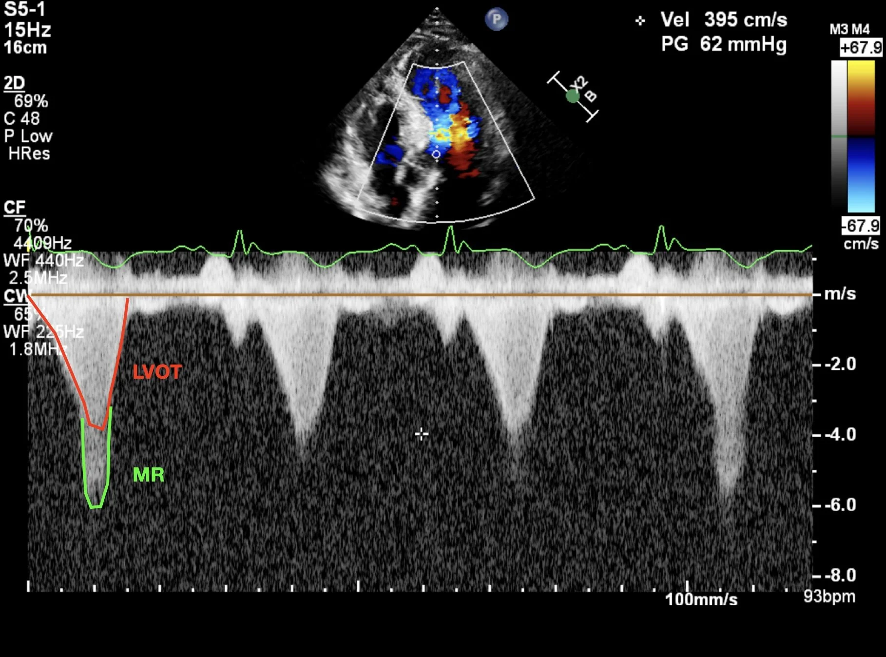
Review this patient’s imaging - this is CW doppler through the LV outflow tract.
Note that on the first and last beats an underlying MR trace is picked up with a merged signal (red is LVOT, green is MR). We know this because the MR trace is more symmetrical and crucially it has a Vmax of 6m/s thus cannot be the LVOT velocity. The true outflow tract doppler trace is seen in the middle beats, and the Vmax is just under 4m/s (giving a gradient of 62mmHg which is very significant)
Management of Dynamic LVOTO
Dynamic LVOTO is scary and counterintuitive to manage but once identified the general management principles are as follows:
1) Treat the underlying pathology
Source control of sepsis, correct hypovolaemia or bleeding.
2) Optimise preload
Adequate preload is important to fill the LV and splint open the LVOT.
Rapid fluid boluses of 250-500ml with serial echo assessment and pause any ongoing diuresis.
3) Increase afterload
Whilst counter-intuitive, increasing aortic pressure will help to splint open the LVOT and reduce dynamic obstruction. Pure vasopressors are often first line here as they will increase PVR and cause a reflex decrease in heart rate without increasing inotropy. Consider:
Phenylephrine (one of the few times when this is a good choice of vasopressor)
Vasopressin will increase PVR without increasing inotropy
Noradrenaline and metaraminol are also reasonable choices, and usually more familiar.
4) Reduce contractility
Avoid (or cease) any inotropes such as adrenaline or dobutamine as increasing LV contractility will just worsen ongoing LVOTO. If an intra-aortic balloon pump is in situ, consider pausing it to allow afterload to increase.
5) Maintain sinus rhythm and slow the heart rate
Aim for a low-normal heart rate in the range of 55-75bpm to optimise diastolic filling and preload. Arrythmias such as AF with RVR may not be well tolerated, and if so will need to be aggressively treated.
If there is ongoing tachycardia consider cautious beta-blockade to control heart rate. One option is Esmolol due to its short half life. It can be rapidly ceased if it worsens haemodynamic compromise
6) Expert advice and mechanical supports
In extremis VA ECMO can effectively support a patient with dynamic LVOTO but if the issue is identified early then the interventions above are usually sufficient.
Very rarely dynamic LVOTO can be driven by RV failure due to RV dilation and ventricular interdependence compressing the LVOTO. In this scenario seek expert advice, and tailor your management to supporting the RV as best possible without worsening the LVOTO.
Case Progress:
The patient had their dynamic LVOTO identified early and responded well to treatment.
They were discharged on day 3, on dual anti-platelets, bisoprolol, and dapagliflozin.
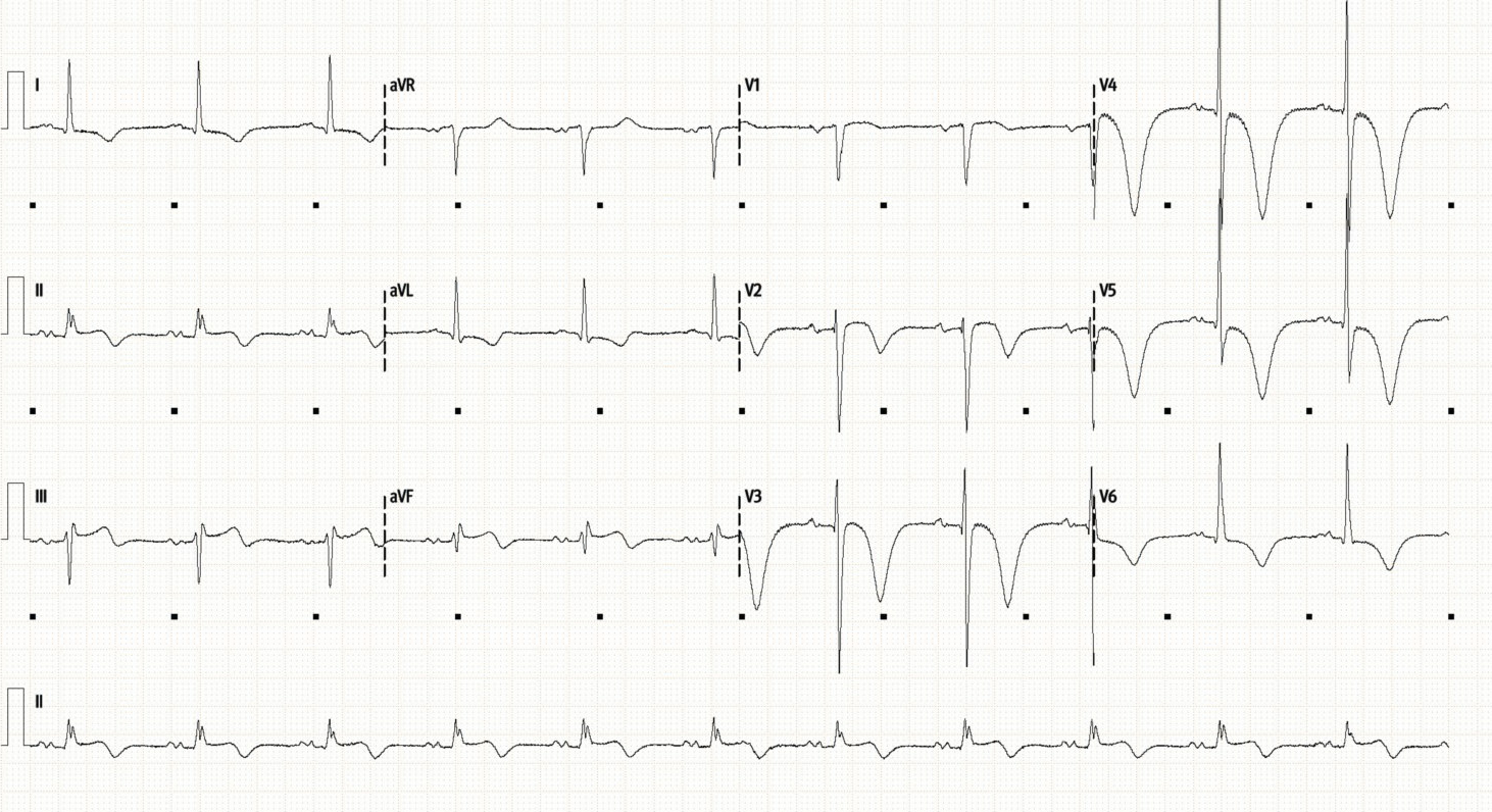
Their ECG on discharge is shown below - note the marked T-wave inversion throughout the precordial leads.